Eye Care Center is dedicated to providing the absolute most cutting edge technology available. Dr. Belinda Dobson introduces new technology into our patient care process annually. Next time you visit us, you are certain to be surprised with the newest in technology advancements.
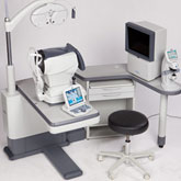
Out with the old, in with the new! At Eye Care Center, Dr. Belinda Dobson uses the TRS-5100, which is Marco's latest generation of digital refraction technology.
Replacing the standard refractor, it allows Dr. Belinda Dobson to control the entire refraction process from a keypad. Do you become anxious about picking from “Which is better, one or two?” With our new technology, this becomes effortless and obsolete. It uses dual imagining so you never have to ‘flip back and forth’ between options! It’s automated and digital, and more accurate than ever before. Come see what you have been missing. See more precise. See more clearly. See the difference.
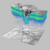
This new high-performance OCT instrument from Carl Zeiss Meditec offers a technological quantum leap forward. Featuring spectral domain technology, Cirrus HD-OCT delivers exquisite high-definition 3-D images of the ocular structures. High Definition data acquisition and advanced analysis provide precise registration and excellent reproducibility critical for glaucoma detection and management.
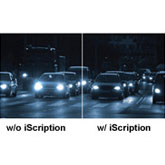
For the first time, experience precision optics with the absolute most high definition lenses available. Experience better night/low-light vision. For example: Looking directly at a light source at night such as car lights, results in glare and halo effects. These are reduced by i-Scription technology.
Experience Better visual contrast. For example: See improved contrast when observing white letters on a black background, which is especially challenging for the eyes. i-Scription technology by Zeiss sharpens contrast.
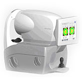
The iProfiler is an instrument designed to determine the exact ‘fingerprint’ of your eyes. This makes it possible to produce Zeiss customized lenses using i.Scription and deliver your best vision possible. Around 1,500 measuring points are used to determine a precise visual profile of your eye. Even the slightest deviations from the ideal shape of the eye can impact your vision. Based on your possible visual profile, we can determine your best prescription possible.
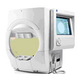
Validated by decades of research and clinical experience, HFA is the accepted standard of care in glaucoma diagnosis and management. HFA has the widest range of testing protocols on any perimeter. The ergonomic design promotes maximum comfort and patient access. It also features screening options that allow Dr. Dobson to screen patients for potential vision problems as part of a thorough eye health assessment.
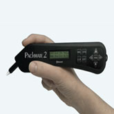
A pachymeter is a medical device that utilizes ultrasound technology to measure the thickness of the eye’s cornea. It is used to perform corneal pachymetry prior to LASIK surgery, for Keratoconus screening, and is useful in screening for patients suspected of developing glaucoma, as well as other corneal diseases. At Eye Care Center, we also utilize pachymetry for the management of an eye condition called Fuch's corneal dystrophy.
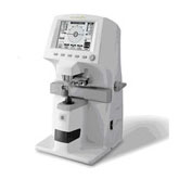
Eye Care Center utilizes lensometry to compare your new prescription to your old glasses. Often patients want to know if their prescription has changed but forget to bring in their old prescription scripts. Lensometry obtains a reading on your old glasses to quantify the changes in refractive error.
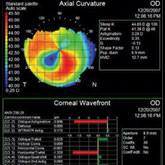
The cornea is the front structure of the eye which serves as the window for your vision. Many times with conditions such as corneal astigmatism, it is difficult to properly fit specialty contact lenses without a more in-depth analysis of the cornea.
Corneal topography allows Dr. Belinda Dobson to map the cornea and successfully fit patients who have previously never been able to wear contact lenses. In addition, corneal topography is used for the diagnosis and management of many eye conditions such as keratoconus, pellucid marginal degeneration, and a host of other rare eye diseases.
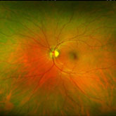
The high definition retinal imaging instrument called Optomap Daytona features state of the art optics which capture a high resolution, panoramic image of your retina, macula, and optic nerve.
The Optomap image facilitates early detection of retinal pathology and other life threatening diseases such as cancer and stroke. The process is quick, painless, and allows your eye doctor to see up to 500% more of the retina than traditional techniques, and often times a dilated pupil exam is not mandatory.


